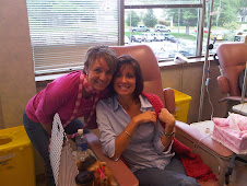
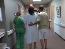
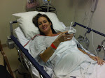
Dr Thiel, radiologist at the Women’s Imaging Center in Burnsville performed an image guided needle aspiration on Mary Jo’s right breast axilli region (armpit) this morning. Mary Jo was a bit nervous before this procedure. It is very similar to the original fine needle aspiration procedure she had performed on the axilliary lymph node which concluded that her breast cancer had spread to her lymph nodes. Your armpit is a tender area to begin with and then to have what looks like a knitting needle stuck into you is all a bit unnerving.
The procedure went well however the doctor was not able to reasonably drain what had been assumed to be a “fluid filled void”. The doctor made several image guided attempts to aspirate but lump and its contents appear to be more consistent with a solid than a fluid. The medical staff has no explanation at this time and we will all have to await the pathology report on the small quantity of fluid that Dr Thiel was able to aspirate from the lump before we know conclusively what the lump contains.
Dr Thiel cautioned Mary Jo that the pathology lab results would not likely be available until Monday but he also tried to calm our obvious anxiety by adding that he doesn’t think this is cancer. Dr Thiel said cancer typically appears a whitish shadowy image on an ultrasound and Mary Jo’s lump appears as a dark spot, which is typically consistent with fluid.
We continue pray that this is not more cancer and just anxiously wait for the pathology report. The waiting is the hard part.

.jpg)


.jpg)

.jpg)
.jpg)


.jpg)


No comments:
Post a Comment